Compact Bone Diagram Microscope : Lab 1.5 - Structures of Compact Bone (Microscope) - YouTube. The spaces between the trabeculae contain red or yellow marrow, depending on a person's age and on which bone it is. Bone tissue anatomy tissue components at right angles to the central canal. Compact bone is formed in concentric circles. The basic microscopic unit of bone is an osteon (or haversian system). This video describes the microscopic anatomy of compact bone.
A diagram of the anatomy of a bone, showing the compact bone. Compact bone is very strong, and it provides the supportive strength of long bones. A structural unit of compact bone consisting of a central canal surrounded by concentric cylindrical lamellae of matrix. The larger ovals are blood vessels running through the bone. This human bone section shows the haversian canal (or osteon) structure of compact bone tissue.
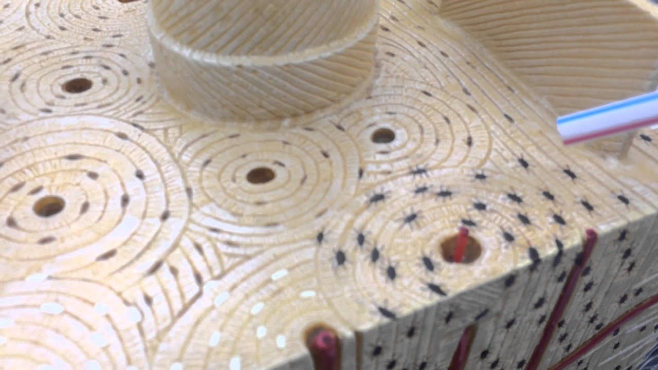
MICROSCOPIC STRUCTURES OF COMPACT BONE (WEDGE OF BONE) - YouTube from i.ytimg.com Bone tissue is one of the main components of the skeletal system (other components include bone marrow/marrow cavity, collagen fibers etc). Compact bone, or cortical bone, mainly serves a mechanical function. It can be found under the periosteum and in the diaphyses of long bones, where it provides support and protection. A structural unit of compact bone consisting of a central canal surrounded by concentric cylindrical lamellae of matrix. Compact bone, also called cortical bone, dense bone in which the bony matrix is solidly filled with organic ground substance and inorganic salts, leaving only tiny spaces (lacunae) that contain the osteocytes, or bone cells.compact bone makes up 80 percent of the human skeleton; This human bone section shows the haversian canal (or osteon) structure of compact bone tissue. Start studying compact bone under microscope. The marrow in these images is red marrow.
Between the rings of matrix, the bone cells (osteocytes) are located in spaces called lacunae.
This is the area of bone to which ligaments and tendons attach. This human bone section shows the haversian canal (or osteon) structure of compact bone tissue. Then, on the photom in the key and draw a bracket enclosing a single osteon the appropriate. Several descriptions of bone structure are given below. Spongy bone is used for more active functions of the bones, including blood cell production and ion exchange. Under the microscope, bone can be divided into two types compact bone forms the outer 'shell' of bone. 114 review sheet 8 microscopic structure of compact bone 11. However, compact bones also serve a function in storing and releasing calcium to the. The compact bone is the main structure in the body for support, protection, and movement. Trabecular bone, also known as cancellous bone or spongy bone, mainly serves a metabolic function. The light spot is a canal that carries a blood vessel and a nerve fiber. Ident fy the structure involved by choosing nenty all st dermcrograph of bone on the right h key and placing its letter in the blank. Compact bone is very strong, and it provides the supportive strength of long bones.
The diagram above shows a longitudinal view of an osteon. Microscopic anatomy of compact bone. The light spot is a canal that carries a blood vessel and a nerve fiber. Decalcified compact bone at 60x magnification. Ident fy the structure involved by choosing nenty all st dermcrograph of bone on the right h key and placing its letter in the blank.
How do compact and spongy bones differ? - Quora from qph.fs.quoracdn.net The cells of compact bone, which is also called cortical bone, appear to be tightly packed into a solid mass. The darker ring consists of layers of bone matrix made by cells called. Bone tissue anatomy tissue components at right angles to the central canal. A structural unit of compact bone consisting of a central canal surrounded by concentric cylindrical lamellae of matrix. There are pores and spaces even in compact bone. Compact bone, or cortical bone, mainly serves a mechanical function. It can be found under the periosteum and in the diaphyses of long bones, where it provides support and protection. Compact bone is very strong, and it provides the supportive strength of long bones.
Bone tissue anatomy tissue components at right angles to the central canal.
Start studying microscope structure of compact bone. Several descriptions of bone structure are given below. Each osteon consists of a lamellae of compact bone tissue that surround a central canal (haversian canal). Bone tissue and cells under the microscope introduction. Trabecular bone, also known as cancellous bone or spongy bone, mainly serves a metabolic function. (the inset shows a more highly magnified view.) notice the position of osteocytes in lacunae (cavities in the matrix). The diagram above shows a longitudinal view of an osteon. 5 shows two chips, one a transverse section, the other a longitudinal section, being clamped to microscope slides while the epoxy glue cures. Microscopic structure of a long bone. This human bone section shows the haversian canal (or osteon) structure of compact bone tissue. A structural unit of compact bone consisting of a central canal surrounded by concentric cylindrical lamellae of matrix. Osteons are roughly cylindrical structures that can measure several millimeters long and around 0.2 mm in diameter. Under magnification you can clearly see the system of concentric circles that forms compact bone.
The light spot is a canal that carries a blood vessel and a nerve fiber. (the inset shows a more highly magnified view.) notice the position of osteocytes in lacunae (cavities in the matrix). This human bone section shows the haversian canal (or osteon) structure of compact bone tissue. This type of bone is located between layers of compact bone and is thin and porous. The compact bone is the main structure in the body for support, protection, and movement.
Microscope Slides Exam 1 - Exercise Science 222a with Warren at University of Puget Sound ... from classconnection.s3.amazonaws.com Bone tissue and cells under the microscope introduction. It is thick and dense. Next, the bone slice, like the one in fig. It is lined by the endosteum and is filled with yellow bone marrow. The cells of compact bone, which is also called cortical bone, appear to be tightly packed into a solid mass. Bone tissue is one of the main components of the skeletal system (other components include bone marrow/marrow cavity, collagen fibers etc). Compact bone, or cortical bone, mainly serves a mechanical function. Under magnification you can clearly see the system of concentric circles that forms compact bone.
Learn vocabulary, terms, and more with flashcards, games, and other study tools.
This is the area of bone to which ligaments and tendons attach. It is lined by the endosteum and is filled with yellow bone marrow. There are small canals that run through the bone, which allow blood vessels to penetrate it. There are pores and spaces even in compact bone. A structural unit of compact bone consisting of a central canal surrounded by concentric cylindrical lamellae of matrix. The cells of compact bone, which is also called cortical bone, appear to be tightly packed into a solid mass. Ident fy the structure involved by choosing nenty all st dermcrograph of bone on the right h key and placing its letter in the blank. The remainder is cancellous bone, which has a spongelike appearance with numerous large spaces and is found in the. Decalcified compact bone at 60x magnification. However, compact bones also serve a function in storing and releasing calcium to the. Long bone, compact bone and spongy bone The compact bone is the main structure in the body for support, protection, and movement. The marrow in these images is red marrow.
The basic microscopic unit of bone is an osteon (or haversian system) compact bone diagram. Osteons are roughly cylindrical structures that can measure several millimeters long and around 0.2 mm in diameter.
Compact Bone Diagram Microscope : Lab 1.5 - Structures of Compact Bone (Microscope) - YouTube
Reviewed by PIKACHU
on
Agustus 12, 2021
Rating: 5

 Reviewed by PIKACHU
on
Agustus 12, 2021
Rating:
Reviewed by PIKACHU
on
Agustus 12, 2021
Rating:


Post a Comment